Can Post-Mortem Human Brain Tissue Control a Robot?
A new preprint wired adult cortical slices from autopsy tissue to a robotic hand. Some slices learned all three note associations, but the robot did much of the work.

Nil Ni is a seasoned journalist specializing in emerging technologies and innovation. With a keen eye for detail, Nil brings insightful analysis to Stanford Tech Review, enriching readers’ understanding of the tech landscape.
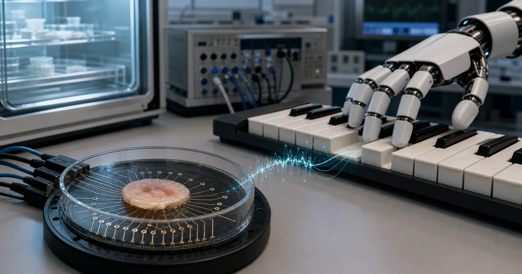
A small piece of adult human cortex, collected during an autopsy, sat on a bed of electrodes in Montpellier. On the other side of the interface was a robotic hand above three piano keys. After three days of paired stimulation, sound input could make the tissue produce an electrical pattern that software translated into the matching finger movement.
That is the striking core of a new Research Square preprint. It is also much narrower than the viral descriptions of a dead brain waking up inside a robot, or of The Matrix becoming real.
The study is best understood as an experiment in associative plasticity. It asks whether preserved adult human cortical tissue can form several durable links between inputs and outputs. The robot makes those links visible, but the tissue is not playing music, choosing a tune, or showing evidence of consciousness.
My verdict: this is a convincing demonstration of durable associative plasticity in adult human cortical tissue, but a weak demonstration of autonomous robotic intelligence. The result could matter more as a new laboratory model for neuroscience and drug testing than as a prototype biological computer.
What the researchers actually built
The team, led by researchers at France's CNRS and the University of Montpellier, used organotypic post-mortem adult brain explants, abbreviated OPABs. According to the paper, the samples came from the primary motor cortex of three donors whose post-mortem interval was under 12 hours. The protocol had ethics approval, prosecutor authorization, and family non-opposition.
Researchers cut the tissue into slices 300 micrometers thick and roughly 5 millimeters across, then kept the slices at an air-liquid interface in controlled culture conditions. This was not an intact brain. It was a thin section of cortex and white matter containing living cells and local circuitry.
The tissue rested on a 60-electrode microelectrode array. The team selected three electrodes as motor outputs and three as sensory inputs. Each motor channel was connected to one finger of a Shadow Robot hand. Those fingers pressed the notes LA, TI, and DO on a keyboard. A microphone and sound-decoder program identified each note and routed a stimulation back to the corresponding sensory electrode.
The preparation therefore had a minimal body: three possible actions and three possible auditory inputs. It also had a substantial digital support system. One computer recorded and stimulated neural activity. A C++ controller converted a selected output channel into a finger movement. A C# decoder converted microphone audio into one of three note labels.
How the tissue was trained
On a priming day, the researchers stimulated all three motor electrodes, followed 12 milliseconds later by all three sensory electrodes. For the next three days, the motor channels were activated in a blocked random pattern. Each activation moved a robotic finger, which pressed a key. The decoded sound was then paired with the next activation of that same motor channel, preserving the 12-millisecond interval used to induce synaptic plasticity.
Each tissue slice received 300 training activations per day. In some cases, the paper says, the team simulated the robot movement and note decoding in software during training for logistical reasons. The biological event under study was the timed pairing of electrical activity, not the physical experience of a hand moving through space.
Calling this “unsupervised learning” is technically defensible because the tissue received no reward signal or correction telling it whether a response was right. In ordinary language, however, the phrase is easy to misread. The associations were engineered through an explicit stimulation schedule. The tissue did not discover a keyboard, decide which sounds mattered, or teach itself a task from an open environment.
On Day 4, the team also calibrated the readout. For each sensory input, researchers stimulated the tissue once and selected the strongest responding electrode within a 3-by-3 neighborhood around the trained motor electrode. That peak electrode then controlled the corresponding finger. This step is reasonable engineering, but it means the robot's output depended on a post-training decoder chosen with knowledge of the intended mapping.
The result is real, but modest
Sixteen explants were included in the main imitation analysis. Before training, mean accuracy across the three possible notes was 31.22 percent, close to the 33.3 percent expected by chance. On Day 4, mean accuracy reached 58.5 percent. Ten of the 16 explants correctly learned at least two of the three note associations, and three achieved perfect performance across all three notes in their test sessions.
The experiment's most useful headline is a 27.3-percentage-point gain from Day 1 to Day 4, not a dead brain playing piano.
Several controls make the learning interpretation stronger. When the same sensory inputs were paired with randomly chosen, untrained output electrodes, Day 4 accuracy averaged 36.6 percent. The trained and untrained paths diverged significantly over time. Blocking AMPA and NMDA receptor activity with CNQX and AP-V reduced performance in six trained explants, supporting the conclusion that neural transmission rather than a software artifact carried the association.
The researchers also infected four high-performing explants with Tahyna virus and compared their Day 7 results with seven healthy explants. Performance was lower after infection. In a separate longitudinal observation, four of five explants retained learned associations for at least 17 days.
These findings extend the team's peer-reviewed 2024 work in Brain Stimulation, which showed that similarly prepared adult human brain explants could retain synaptic plasticity after death and develop changed responses after multiday paired stimulation. The new study adds multiple parallel associations and a physical output device.
How much of the robot's behavior came from the tissue?
The answer is: an important choice signal, surrounded by conventional software.
During a test, a person played a sequence of 10 to 18 notes. For every note, the system stimulated the matching sensory electrode five times. It averaged each candidate motor electrode's response over one-second windows, subtracted a pre-training baseline, and selected the largest remaining response. The robot controller then moved the finger already assigned to that channel.
The explant supplied the relative electrical response that selected one of three outputs. It did not decode sound, control joints, plan timing, or evaluate its own errors. Those jobs belonged to the microphone decoder, the analysis pipeline, and the robot controller.
This division of labor does not invalidate the experiment. Every brain-computer interface needs signal processing and actuation layers. It does change the headline. “Human cortical tissue formed three reversible associations that a decoder used to select robot fingers” is less cinematic than “dead brain controls robot,” but it is more accurate.
Why this is not a brain in a vat
The brain-in-a-vat thought experiment requires a mind receiving a complete simulated world while remaining unaware of its true physical situation. Nothing in this study approaches that condition.
The explants were tiny cortical slices, not whole brains. They lacked the distributed architecture that links cortex with thalamus, brainstem, body regulation, memory systems, and integrated senses. The experiment measured local electrical plasticity and task performance. It did not test subjective experience, self-awareness, pain, identity, or memory from the donor.
The paper itself does not claim consciousness or sentience. That restraint matters because this field has already fought over anthropomorphic language. A 2022 system called DishBrain showed that cultured neurons could adapt within a simulated Pong environment, but its use of “sentience” prompted a published response from 26 neuroscientists arguing that the evidence did not justify the term. The safer rule is simple: learning is a measurable change in performance; consciousness is a separate claim requiring separate evidence.
That does not eliminate ethical questions. Human neural tissue linked to machines deserves careful oversight, especially as preparations become larger, more connected, or more richly stimulated. It does mean that today's evidence supports a precautionary conversation, not a declaration that a person survived in a dish.
What the preprint still needs to prove
The study is under review and has not yet passed journal peer review. Its main result rests on 16 explants from only three donors. Explants from the same person are not fully independent biological replicates, yet the reported analysis treats slices as the experimental units and does not present donor-stratified effect sizes. An independent replication across more donors would show whether the effect is robust to age, cause of death, post-mortem interval, and tissue condition.
Future work should lock the decoder before testing, report performance both with and without the Day 4 peak-electrode calibration, and expand beyond three choices. A three-note task gives chance performance of one in three and leaves little room to measure how capacity scales. A ten-note task, novel sequences, and delayed tests would better separate general associative memory from a small, engineered lookup table.
The long-term result is promising but preliminary. Four retained explants at Day 17 are a signal worth following, not yet a reliable estimate of memory duration. Likewise, the virus experiment could become a useful disease model, but four infected samples are too few to support broad conclusions about cognition after viral infection.
The real breakthrough may be a better test bench
The robot hand attracts attention because it turns invisible electrophysiology into an action anyone can understand. But the practical value may lie elsewhere.
Adult human cortical tissue preserves mature cell types and local organization that stem-cell-derived organoids do not fully reproduce. If researchers can induce and measure repeatable associations in that tissue, they gain a human experimental system for studying how drugs, infections, and disease processes alter learning-related circuitry. The paper's receptor-blocking and virus experiments are early examples of that use.
Biological computing is the more speculative branch. The current preparation needs careful culture, repeated stimulation, custom decoding, and a computer-controlled interface to choose among only three outputs. Silicon can solve this particular task more cheaply and reliably. The case for hybrid computing will require a task where living tissue provides a measurable advantage in energy, adaptability, or sample efficiency, not just novelty.
For now, the experiment has crossed a meaningful line without crossing the science-fiction one. Post-mortem adult human cortical tissue can retain trainable associations and can provide a control signal to a robot days later. That is enough to take seriously. It is not enough to call the tissue a mind, a pianist, or the first resident of The Matrix.
Cover image generated by Stanford Tech Review.